Pushing the envelope of flow cytometry via multipass-imaging
Microscopy has long been a cornerstone in modern medicine and biology. From its inception in the 17th century to the present, it has played an indispensable role in our exploration of the natural world. It is essential to recognize that traditional light microscopy encounters challenges when imaging objects with weak absorption characteristics, such as biological cells.
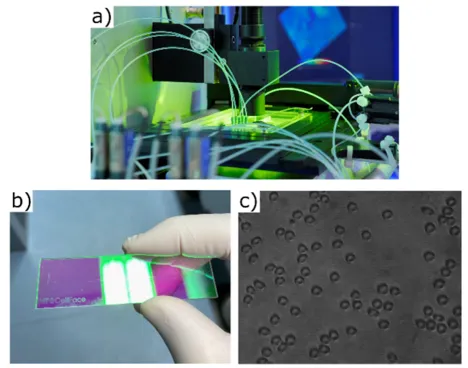
Together with our collaboration partners at the Heinz-Nixdorf Chair of Biomedical Electronics at the Translational Cancer Research Center (TranslaTUM), we have developed low-profile multipass cytometry chips integrated into a high-throughput microfluidic channel. These chips are designed for implementation in any in-vitro diagnostic clinical setting, enabling contrast enhancement in conventional light microscopy as well as phase contrast microscopy of biological specimens. This enhancement is achieved by multipassing a coherent light source through Fabry-Perot micro-cavities.
Variable flow conditions and instabilities often affect cytometric measurements, potentially leading to mode-hopping and loss of contrast enhancement. In our most recent adaptation, we employ an ultra-low bandwidth λ-tunable laser source as the chosen imaging light source, ensuring that resonance conditions are met. All these measures combined not only streamline the measurement of microscopic biological specimens but also provide improved visibility and adaptability required for various applications in a changeable clinical setting.
First measurements with the novel design of the multi-pass chips on human red blood cells (HRBC) were recently performed at the chair and are currently being evaluated. In addition, we are working on the development of a comprehensive automated cell registration and analysis framework adapted for the experimental challenges arising in multipass-microscopy, allowing rapid data processing.
As we aim to harness the advantageous characteristics of both multipassing and phase-sensitivity in measurements, our efforts are also directed towards incorporating multipass-imaging into Digital Holography Microscopy (DHM). This advancement holds great potential for enhancing cytometry techniques when dealing with low-contrast specimens. For this purpose, we are currently building and testing a patented novel design of a shearing-based common-path microscope.