Eggl, E., Schleede, S., Bech, M., Achterhold, K., Loewen, R., Ruth, R.D. & Pfeiffer, F. X-ray phase-contrast tomography with a compact laser-driven synchrotron source. Proc Natl Acad Sci USA 112, 5567–5572 (2015) [http://doi.org/10.1073/pnas.1500938112]
Purpose: Here we present, to our knowledge, the first phase-contrast tomography acquired at a compact light source, a recently developed compact synchrotron based on inverse Compton scattering.
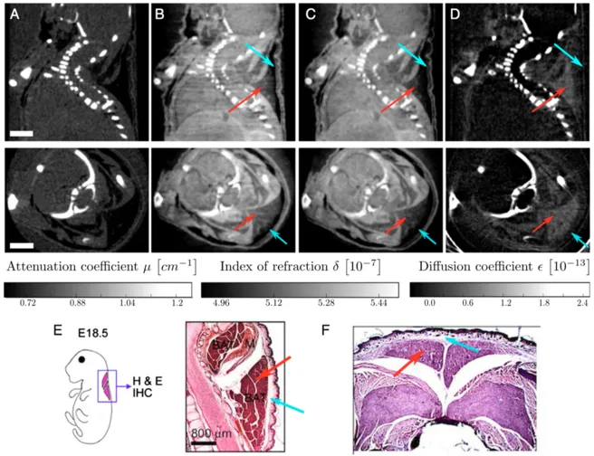
Figure: Reconstructed slices of a grating-based, multimodal CT scan of a biological sample (a formalin fixated infant mouse). Shown are sagittal (Top Row) and axial (Bottom Row) slices. The images show that brown adipose tissue is visible and can be discriminated from white adipose tissue in phase contrast (B and C) and dark-field contrast (D), but not in absorption contrast (A). (Scale bar, 2 mm.) Histological slices presented in E and F support this claim. E shows a sagittal section of the cervical/thoracic area stained with H&E of a mouse embryo . F shows an axial section of the interscapular area stained with H&E of a mouse embryo . Red arrows indicate brown adipose tissue; blue arrows indicate white adipose tissue.