The SmartX project has been awarded with an ERC Synergy Grant from the renowned European Research Council, focusing on groundbreaking future technology: developing ultra-detailed, low-radiation X-ray imaging
SmartX
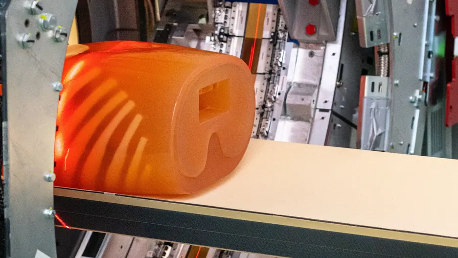
The SmartX project is researching more detailed imaging procedures in order to diagnose lung diseases better and earlier. The focus is on chronic obstructive pulmonary disease (COPD). Conventional X-rays and CT scans are often inadequate for the early detection of COPD and are associated with high radiation doses. The aim is to develop a new type of detector for the dark-field X-ray procedure that requires 50 percent less radiation dose compared to the already low-radiation procedure. An X-ray detector is the counterpart to the radiation source and produces the X-ray image. While conventional X-rays are based on the attenuation of the X-ray light, the dark-field X-ray developed at TUM uses the so-called small-angle scattering of the X-ray light. This allows additional information to be obtained about the nature of the microstructure of the lung tissue.
SmartX is led by Franz Pfeiffer, Professor of Biomedical Physics and Director of the Munich Institute of Biomedical Engineering at TUM, together with José M. Benlloch Baviera, Professor at the CSIC in Valencia, Edoardo Charbon, Professor at the EPF de Lausanne, and Daniela Pfeiffer, Professor of Radiology at TUM.
Patient study demonstrates benefits of dark-field X-ray technology
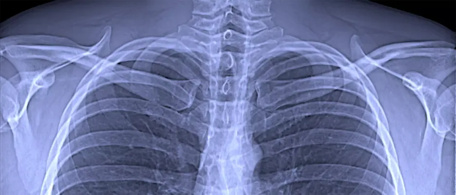
A research team at the Technical University of Munich (TUM) has, for the first time, produced dark-field X-ray images of patients infected with the corona virus. In contrast to conventional X-ray images, dark-field images visualize the microstructure of the lung tissue, thereby providing additional information. This approach has the potential to provide an alternative to computed tomography (CT), which requires a significantly higher radiation dose.
Prototype of a clinical CT device combines dark-field X-ray and conventional technology

For the first time, a team of researchers at the Technical University of Munich (TUM) has integrated the dark-field X-ray method into a CT scanner suitable for clinical use. Dark-field imaging provides additional information to conventional X-ray imaging. With the new prototype, it is now possible to produce three-dimensional dark-field X-ray images.
Dark-field X-ray technology improves diagnosis of pulmonary ailments

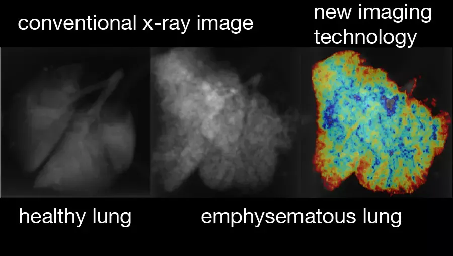
For the first time, researchers at the Technical University of Munich (TUM) have successfully used a new X-ray method for respiratory diagnostics with patients. Dark-field X-rays visualize early changes in the alveolar structure caused by the lung disease COPD and require only one fiftieth of the radiation dose typically applied in X-ray computed tomography. This permits broad medical application in early detection and treatment follow-up of respiratory ailments.
Low-dose radiographs could reveal typical lung changes
Researchers at the Technical University of Munich (TUM) have developed an innovative x-ray method for lung diagnostics, which they now plan to test in one of its first applications for diagnosis of the respiratory ailment Covid-19 caused by Coronavirus. The method could clearly identify abnormalities typical of the illness and involves a significantly lower radiation dose than the computed tomography methods currently in use. Last week, the German Federal Office for Radiation Protection (BfS) issued the approval necessary for the tests.
New staining method enables Nano-CT imaging of tissue samples

To date, examining patient tissue samples has meant cutting them into thin slices for histological analysis. This might now be set to change – thanks to a new staining method devised by an interdisciplinary team from the Technical University of Munich (TUM). This allows specialists to investigate three-dimensional tissue samples using the Nano-CT system also recently developed at TUM.
X-rays from the MuCLS compact synchrotron open up new possibilities for heart examinations

The most prevalent method for obtaining images of clogged coronary vessels is coronary angiography. For some patients, however, the contrast agents used in this process can cause health problems. A team at the Technical University of Munich (TUM) has now demonstrated that the required quantity of these substances can be significantly reduced if monoenergetic X-rays from a miniature particle accelerator are used.
Novel imaging device creates high-resolution 3D-x-rays of tiny velvet worm legs

Computer Tomography (CT) is a standard procedure in hospitals, but so far, the technology has not been suitable for imaging extremely small objects. In PNAS, a team from the Technical University of Munich (TUM) describes a Nano-CT device that creates three-dimensional x-ray images at resolutions up to 100 nanometers. The first test application: Together with colleagues from the University of Kassel and Helmholtz-Zentrum Geesthacht the researchers analyzed the locomotory system of a velvet worm.
Leibniz Prize winner Franz Pfeiffer to head TUM Integrative Research Center

Two years after its founding at the Technical University of Munich (TUM), the Munich School of BioEngineering (MSB) is expanding its scope of action: The upcoming opening of the MSB-associated Central Institute for Translational Cancer Research (TranslaTUM) will give physicists, engineers and physicians a shared new laboratory building for transdisciplinary research in the midst of the TUM’s Klinikum rechts der Isar in Munich. In addition, a new structure for the Bavarian NMR Center (BNMRZ) will soon open on the Garching campus. The new building for protein research, expected to be finished in two years and also located in Garching, completes the most topically diverse bio-engineering structure in all of Europe. The new construction represents an investment of more than 135 million Euros, half of which is being financed by the German Federal Government. Leibniz Prize winner Prof. Franz Pfeiffer (44) will succeed the founding director Prof. Axel Haase as of April 1, 2017.
Nine ERC Grants for research

Nine scientists from the Technical University of Munich (TUM) won out in the latest round of ERC grants. The projects receiving funding are in the disciplines Medicine, Physics and Informatics and deal with a highly varied range of topics such as investigation of autoimmune diseases, innovative algorithms and bio-nanotechnology.
New X-ray method uses scattering to visualize nanostructures
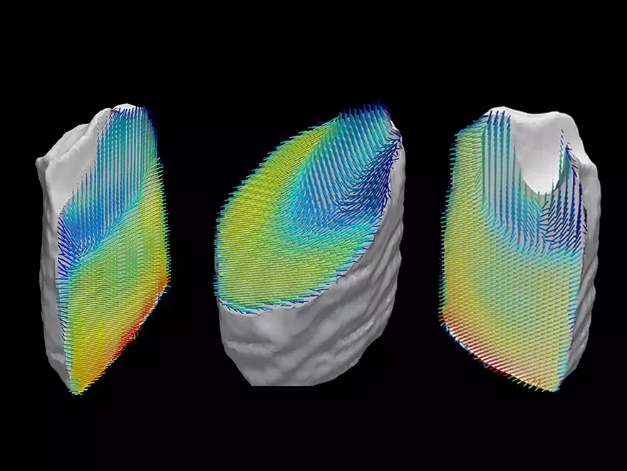
Both in materials science and in biomedical research it is important to be able to view minute nanostructures, for example in carbon-fiber materials and bones. A team from the Technical University of Munich (TUM), the University of Lund, Charité hospital in Berlin and the Paul Scherrer Institute (PSI) have now developed a new computed tomography method based on the scattering, rather than on the absorption, of X-rays. The technique makes it possible for the first time to visualize nanostructures in objects measuring just a few millimeters, allowing the researchers to view the precise three-dimensional structure of collagen fibers in a piece of human tooth.
New X-ray method for 3D-images of soft tissue developed
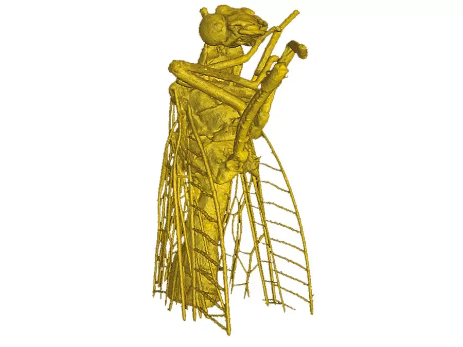
When the physicists Prof. Stefan Karsch, researcher at the Max Planck Institute for Quantum Optics and Ludwig-Maximilians-Universität München, and Prof. Franz Pfeiffer, TUM professor for Biomedical Physics, illuminate a tiny fly with the new technique, the resulting image captures even the finest hairs on the wings of the insect. For the first time, the scientists have coupled their technique for generating X-rays from laser pulses with phase-contrast X-ray tomography to visualize tissues in organisms. The result is a three-dimensional view of the insect in exact detail.
Novel X-ray technology improves contrast in soft tissue
Soft tissue disorders like tumors are very difficult to recognize using normal X-ray machines. There is hardly any distinction between healthy tissue and tumors. Researchers at the Technische Universität München (TUM) have now developed a technology using a compact synchrotron source that measures not only X-ray absorption, but also phase shifts and scattering. Tissue that is hardly recognizable using traditional X-ray machines is now visible.
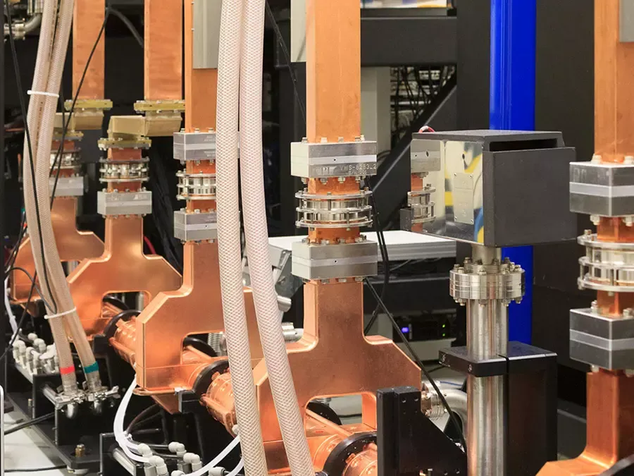
World’s first mini particle accelerator for high-brilliance X-rays at TUM
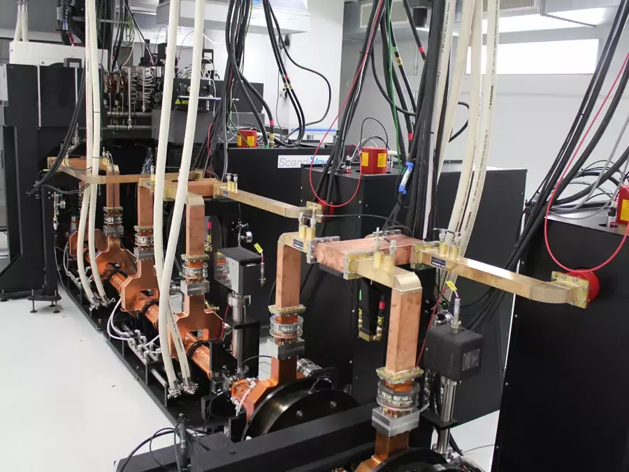
For some years now it has been possible to generate high-brilliance X-rays using ring-shaped particle accelerators (synchrotron sources). However, such installations are several hundred meters in diameter and cost billions of euros. The world’s first mini synchrotron was inaugurated today at Technical University of Munich (TUM). It can generate high-brilliance X-rays on a footprint measuring just 5 x 3 meters. The new unit will be used chiefly to research biomedical questions relating to cancer, osteoporosis, pulmonary diseases and arteriosclerosis.
Imaging method improved by scrambling X-rays from a new source
X-ray phase-contrast imaging can provide high-quality images of objects with lower radiation dose. But until now these images have been hard to obtain and required special X-ray sources whose properties are typically only found at large particle accelerator facilities. Using a laboratory source with unprecedented brightness, scientists from the Technische Universität München (TUM), the Royal Institute of Technology in Stockholm (KTH) and University College London (UCL) have demonstrated a new approach to get reliable phase contrast with an extremely simple setup.
Prototype represents a step toward enhanced soft-tissue tomography

A promising approach for producing medical images with enhanced soft tissue visibility — grating-based x-ray phase contrast—has now advanced from bench-top studies to implementation in an in vivo preclinical computed tomography scanner. A German, Swedish, and Belgian team led by scientists at the Technische Universität München (TUM) published the first experimental results demonstrating the practical potential of this technology, which can significantly improve the contrast in CT scans. This work, reported in the Proceedings of the National Academy of Sciences, could mark a critical step in moving beyond proof-of-concept experiments to applications —including in vivo preclinical imaging with small-animal models in the mid-term future and, in the long term, medical CT scanning.
X-ray technology improves early detection of pulmonary disease
Severe lung diseases are among the leading causes of death worldwide. To date they have been difficult to diagnose at an early stage. Within an international collaboration scientists from Munich now developed an X-ray technology to do just that. Now they are working on bringing the procedure into medical practice.
German Research Foundation awards top science prize to TUM physicist:

Physicist Franz Pfeiffer is one of the ten winners of the 2011 Leibniz Prize, the German Research Foundation (DFG) has announced. The 38-year-old physicist, holds the Chair for Biomedical Physics at the Technische Universitaet Muenchen (TUM). The price honors his fundamental and applied research in phase-contrast X-ray imaging, which promises significant progress toward early detection of tumors. The Leibniz Prize is Germany's most renowned scientific award and brings each recipient 2.5 million euros in prize money.