Muenzel, D. et al. Spectral Photon-counting CT: Initial Experience with Dual-Contrast Agent K-Edge Colonography. Radiology 283, 723–728 (2017). [https://doi.org/10.1148/radiol.2016160890]
Purpose: To investigate the feasibility of using spectral photon-counting computed tomography (CT) to differentiate between gadolinium-based and nonionic iodine-based contrast material in a colon phantom by using the characteristic k edge of gadolinium.
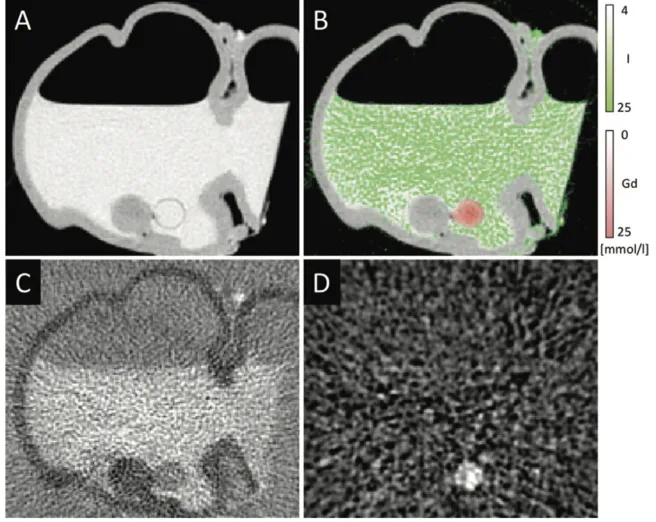
Photon-counting CT of colon phantom. A, Conventional CT scan (window: 2300 HU, level: 1000 HU). B, Conventional CT scan (image in A) with overlay of iodine (green) and gadolinium (red). C, Iodine image (window: 0 mmol/L, level: 80 mmol/L). D, Gadolinium image (window: 5 mmol/L, level: 20 mmol/L). Both material images (C and D) are generated from decomposition algorithm and are visually and quantitatively distinguishable. Therefore, this procedure enables a separation between gadolinium-enhanced polyp tissue and iodine-tagged fluids and feces in colon.