Schaff, F., Bech, M., Zaslansky, P., Jud, C., Liebi, M., Guizar-Sicairos, M. & Pfeiffer, F. Six-dimensional real and reciprocal space small-angle X-ray scattering tomography. Nature 527, 353–356 (2015) [http://doi.org/10.1038/nature16060]
Purpose: Here we present a solution to this problem and obtain a complete SAXS computed tomography, which preserves oriented scattering information.
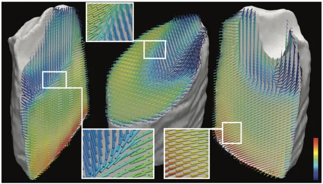
3D visualization of collagen fibre orientation within the tooth sample. The orientation of the coloured bars indicate the mean orientation of collagen fibres obtained from ellipsoid fits to the ratios of scattering intensities of q ranges with and without collagen peaks. The colour represents the average scattered intensity in the collagen range 0.88–0.94 Å−1. The underlying 3D nanomorphology within the entire sample is revealed. An animation showing all slices in one direction is provided in Supplementary Video 2.