Müller, M., de Sena Oliveira, I., Allner, S., Ferstl, S., Bidola, P., Mechlem, K., Fehringer, A., Hehn, L., Dierolf, M., Achterhold, K., Gleich, B., Hammel, H., Mayer, G. & Pfeiffer, F. PNAS 114 (47) 12378-12383 (2017). [https://doi.org/10.1073/pnas.1710742114]
Purpose: We present a laboratory CT device and data processing pipeline to routinely and efficiently generate high-resolution 3D data (≈100 nm) without requiring synchrotron radiation facilities.
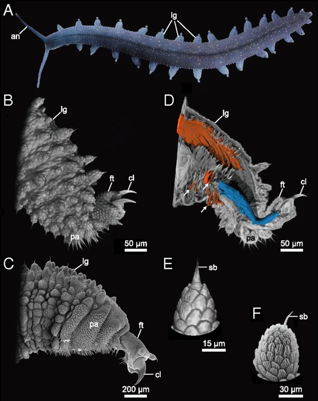
Figure: The onychophoran limb anatomy. Photograph (A), nanoCT volume renderings (B, D, and E), and scanning electron micrographs (C and F) of the onychophoran species E. rowelli. Note that the detail visibility obtained in the volume rendering of nanoCT data from a newborn specimen (B and E) is comparable to that from scanning electron microscopy of an adult specimen (C and F). (A) Overview of a 5-cm-long living specimen walking. Note the repeated arrangement of the legs along the anteroposterior body axis. (B and C) External anatomy of the leg. (D) Digital sagittal section through the same leg as in B showing the leg musculature. The claw retractor muscle is segmented in blue and the leg remotor in orange. Note that this technique allows us to reconstruct single muscle fibers in detail (arrows). (E and F) Detail of a primary papilla of the leg. an, antenna; cl, claw; ft, foot; lg, leg; pa, spinous pad; sb, sensorial bristle.