Gradl, R., Morgan, K. S., Dierolf, M., Jud, C., Hehn, L., Gunther, B., Moller, W., Kutschke, D., Yang, L., Stoeger, T., Pfeiffer, D., Gleich, B., Achterhold, K., Schmid, O., & Pfeiffer, F. Dynamic In Vivo Chest X-ray Dark-Field Imaging in Mice. IEEE transactions on medical imaging, 38(2), 649–656. (2019). https://doi.org/10.1109/TMI.2018.2868999
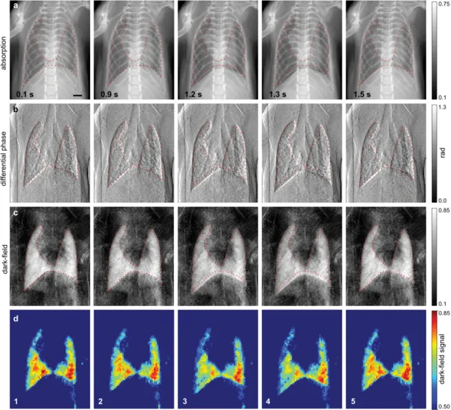
Purpose: In this research project, we demonstrate the first time-resolved dark-field imaging of a breath cycle in a mechanically ventilated mouse, in vivo, which was obtained using a grating interferometer. The measurements show that the dark-field signal depends on the air volume and, hence, the alveolar dimensions of the lung. Conducting this type of scan with animal disease models would help to locate the optimum breath point for single-image diagnostic dark-field imaging and could indicate if the changes in the dark-field signal during breath provide a diagnostically useful complementary measure.
Figure: a) The absorption, b) differential phase, c) dark-field and d) 2×2 spatially-binned dark-field images (shown with a color look-up table), each at five timepoints across the breath cycle.