Scherer, K., Yaroshenko, A., Bölükbas, D. A., Gromann, L. B., Hellbach, K., Meinel, F. G., Braunagel, M., Berg, J. von, Eickelberg, O., Reiser, M. F., Pfeiffer, F., Meiners, S. and Herzen, J. Scientic Reports 1–9 (2017). [http://doi:10.1038/s41598-017-00489-x]
Purpose: Within this letter, we propose to exploit X-ray dark-field imaging as a novel diagnostic tool for the detection of lung cancer on projection radiographs.
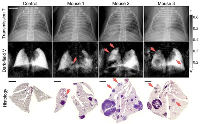
Figure: Typical transmission (top row) and dark-field (middle row)radiograms of a control mouse and three mice with lung tumors, alongside with the corresponding histological sections in H&E staining (bottom row). Small nodules are much easier to detect within the dark-field image (red arrows) than in the attenuation channel, due to a minor impact of overlaying structures and the overall dominant scatter signal of the lung within the thorax.