John, D., Gottwald, W., Berthe, D. et al. X-ray dark-field computed tomography for monitoring of tissue freezing. Sci Rep 14, 5599 (2024). https://doi.org/10.1038/s41598-024-56201-3
Purpose: The potential of X-ray dark-field monitoring for the freezing of tissue models has so far not been investigated in either radiography or computed tomography. In this work, we show that the extent of the frozen region in a porcine phantom mimicking breast tissue can be monitored more accurately with a grating interferometer at a laboratory X-ray source than with attenuation-based imaging alone. For this purpose, the phantom is actively frozen using a pipe connected to a liquid nitrogen reservoir. The imaging results are first demonstrated in radiographs and then transferred to a CT setup.
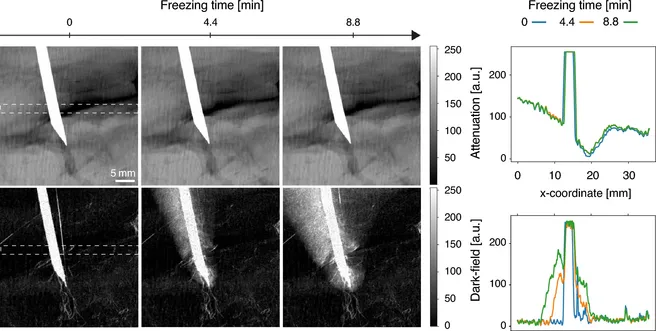
Figure: Series of radiographs (left) and line plots (right) of a pork neck slab frozen using a copper rod connected to a liquid nitrogen reservoir on one end. While the extent of the frozen region is clearly visible in the dark-field images, it is not visually detectable in the attenuation images. The line plots show the signal change over time in the dotted white rectangular region, averaged in the vertical direction.
