Taphorn, K., Kaster, L., Sellerer, T., Hötger, A., & Herzen, J. (2023). Spectral X-ray dark-field signal characterization from dual-energy projection phase-stepping data with a Talbot-Lau interferometer. Scientific reports, 13(1), 767. https://doi.org/10.1038/s41598-022-27155-1
Purpose: Material-selective analysis of spectral X-ray imaging data requires prior knowledge of the energy dependence of the observed signal. Contrary to conventional X-ray imaging, where the material-specific attenuation coefficient is usually precisely known, the linear diffusion coefficient of the X-ray dark-field contrast does not only depend on the material and its microstructure, but also on the setup geometry and is difficult to access. Here, we present an optimization approach to retrieve the energy dependence of the X-ray dark-field signal quantitatively on the example of closed-cell foams from projection data without the need for additional hardware to a standard grating-based X-ray dark-field imaging setup. A model for the visibility is used to determine the linear diffusion coefficient with a least-squares optimization. The comparison of the results to spectrometer measurements of the linear diffusion coefficient suggests the proposed method to provide a good estimate for the energydependent dark-field signal.
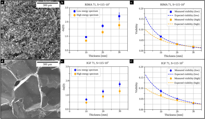
Figure: equired data and results from the optimization. (a,d) Helium-ion microscopy images of RIMA 71 and IGF 71, respectively. (b,e) Measured dark-field signals for increasing thicknesses of the materials. The dark-field values vary for both materials. (c,f) Comparison between measured (data points; standard deviation given as error bars) and expected visibility (dashed line) calculated for the optimized parameters a and b and Eq. (6)