Braig, E.M., Pfeiffer, D., Willner, M., Sellerer, T., Taphorn, K., Petrich, C., Scholz, J., Petzold, L., Birnbacher, L., Dierolf, M., Pfeiffer, F. and Herzen, J. Single spectrum three-material decomposition with grating-based x-ray phase-contrast CT. Physics in Medicine & Biology 65, 185011 (2020) [https://doi.org/10.1088/1361-6560/ab9704]
Purpose: Here, we investigate whether this threefold imaging signal can be used for three-material decomposition at conventional polychromatic x-ray sources operated with more than mean energy. In contrast to the prior approach (Braig et al 2018) and especially to DE-CT, we will evaluate the use of the x-ray dark-field signal in addition to attenuation and phase contrast to simultaneously differentiate between blood coagula, contrast agent and hydroxyapatite clusters.
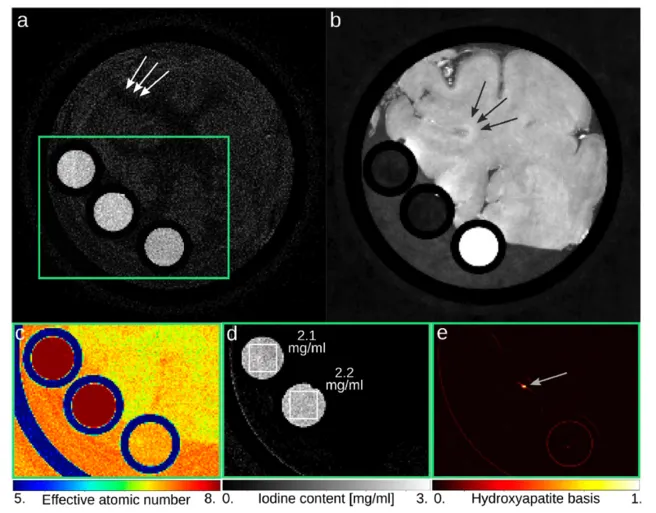
Figure: Multi-modal representation of a part of a pig brain (frontal lobe) based on a single CT acquisition at setup S2. (a) Besides the conventional attenuation image, which is displayed in Hounsfield units ([0; 200] HU), further image representations are available for diagnosis. (b) In the electron density image (displayed in phase Hounsfield units ([−10; 95] HUp)), the clotted blood can clearly be identified and differentiated from the contrast agent. (c) The effective atomic number image is displayed in pseudo colors as known from dual-energy CT. (d) The iodine content can be determined quantitatively by three-material decomposition. (e) The dark-field image is displayed in terms of the normalized hydroxyapatite basis vector and it reveals a calcification within the iodine-filled tube (light gray arrow), which does not occur in the iodine content image. The brain sample was positioned in a plastic tube with 2.9 cm outer diameter. The iodine solutions and the blood coagulum were filled into small plastic tubes with 0.54 cm outer diameter.