Fingerle, A. A., Willner, M., Herzen, J., Münzel, D., Hahn, D., Rummeny, E. J., Noël, P. B. and Pfeiffer, F. Radiology 272(3), pp. 739–748 (2014). [http://doi.org/10.1148/radiol.14130876]
Purpose: To determine if grating-based x-ray phase-contrast computed tomography (CT) can allow differentiation of simulated simple, protein-rich, hemorrhagic, and enhancing cystic renal lesions in an in vitro phantom.
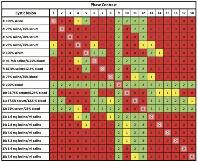
Figure: Graphs show differentiability values for pairwise visual comparison of (a) attenuation-contrast and (b) phase-contrast images of cystic renal lesion phantoms. Numbers 1-18 in column and row headers indicate lesion number. Grading scale for visual differentiability: 0 (red) = differentiation impossible, 1 (yellow) = differentiation uncertain, and 2 (green) = differentiation possible. Highlighted red squares indicate two cyst images of exact same type.