Willner, M., Herzen, J., Grandl, S., Auweter, S., Mayr, D., Hipp, A., Chabior, M., Sarapata, A., Achterhold, K., Zanette, I., Weitkamp, T., Sztrókay, A., Hellerhoff, K., Reiser, M. and Pfeiffer, F. Physics in medicine and biology 59(7), pp. 1557–71 (2014). [http://doi.org/10.1088/0031-9155/59/7/1557]
Purpose: In this study, we investigate quantitative breast tissue characterization using grating-based phase-contrast computed tomography (CT) at conventional polychromatic x-ray sources.
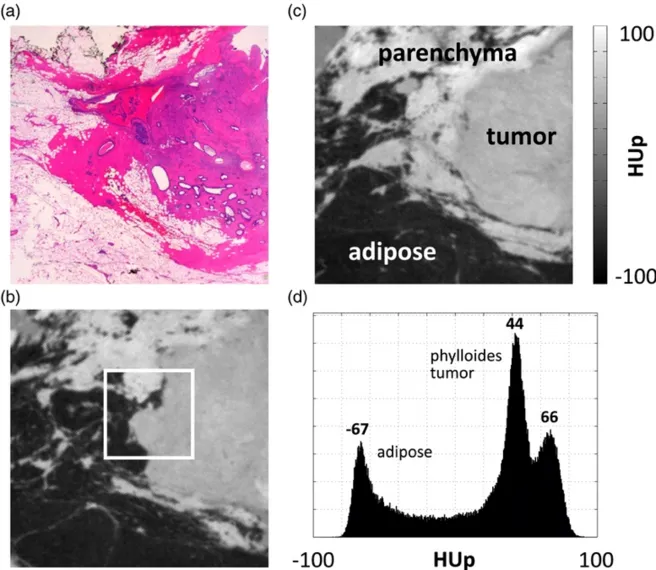
Figure: Histological slice in H&E-staining (a) and two correlated phase-contrast images (b) and (c) of a benign phylloides tumor surrounded by adipose and fibroglandular tissue. The three peaks in the histogram (d) of a cubic region covering all three tissue types resemble the previous results obtained at the synchrotron: adipose tissue between −70 and −65 HUp, tumor tissue around 45 HUp and fibroglandular tissue at about 65 HUp.