Braunagel, M., Birnbacher, L., Willner, M., Marschner, M., De Marco, F., Viermetz, M., Notohamiprodjo, S., Hellbach, K., Auweter, S., Link, V., Woischke, C., Reiser, M. F., Pfeiffer, F., Notohamiprodjo, M. and Herzen, J. Scientific reports. 7, p. 45400 (2017). [<doi:10.1038/srep45400]
Purpose: The present study evaluates the potential of grating-based X-ray phase-contrast computed tomography (gbPC-CT) for visualization and characterization of human RCC subtypes.
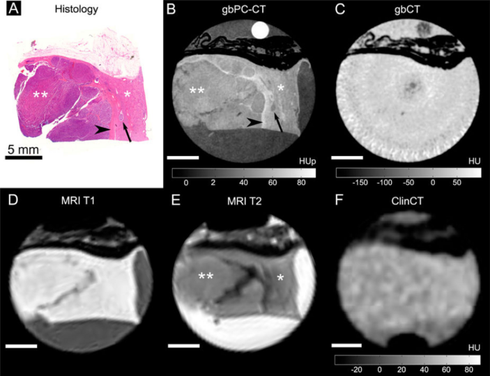
Figure: A perfect match of the histological slice (A) and the phase-contrast CT image (B) was seen with a clear discrimination of the normal cortex (*) with a higher phase-contrast signal and the homogeneous tumor area (**) with lower signal in grating-based phase-contrast CT. Additionally, grating-based phase-contrast CT could depict the pseudocapsule surrounding the tumor with a higher signal (arrowhead) than cortex, a micrometastasis in the cortex (arrow) as well as small linear fibrous strands. In grating-based CT (C) and clinical CT (F), only perirenal fat was visible (hypodense), soft-tissue components could not be differentiated. When correlating grating-based phase-contrast CT (B) with MR images (D,E), a superior discrimination of tumor and normal kidney is seen in the phase-contrast image.