Notohamiprodjo, S., Webber, N., Birnbacher, L., Willner, M., Viermetz, M., Herzen, J., Marschner, M., Mayr, D., Bartsch, H., Saam, T., Auweter, S., Pfeiffer, F., Reiser, M. F. and Hetterich, H., Investigative radiology, 53(1), pp. 26–34 (2017). [http://doi.org/10.1097/RLI.0000000000000408]
Purpose: Grating-based phase-contrast computed tomography (gb-PCCT) relies on x-ray refraction instead of absorption to generate high-contrast images in biological soft tissue. The aim of this study was to evaluate the potential of gb-PCCT for the depiction of structural changes in heart disease.
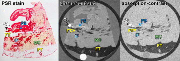
Figure: Myocardial infarction scar (MI). PSR-stained section of the heart sample with MI (left) and corresponding phase-contrast (middle) and absorption-contrast images (right). Myocardium (MC), fatty tissue (FT), fibrotic scars (FS), interstitial fibrosis (IF) and calcification (CL) have been identified as tissue types. Collagen fibers are depicted in red color and myocytes in yellow color in PSR-staining. Strands brighter than MC in phase-contrast images correlate to fibrotic scars in histopathology, but are hardly definable in absorption-contrast images.