Gromann, L. B., De Marco, F., Willer, K., Noël, P. B., Scherer, K., Renger, B., Gleich, B., Achterhold, K., …, Pfeiffer, F. and Herzen, J. ‘In-vivo X-ray Dark-Field Chest Radiography of a Pig.’, Scientific Reports 7(1), 134–7 (2017). [http://doi.org/10.1038/s41598-017-05101-w]
Purpose: In this letter, we present the first in-vivo XDF full-field chest radiographs (32 × 35 cm2) of a living pig, acquired with clinically compatible parameters
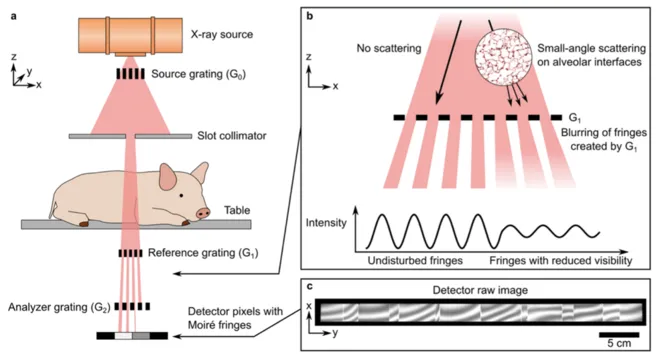
Figure: Full-field X-ray dark-field (XDF) chest radiography scanner. (a) Schematic of the prototype, (b) in case of the lung, millions of micron-sized alveoli (more precisely their air-tissue interfaces) scatter the X-rays, producing the signal, and (c) raw detector image with the fringe pattern.