Grandl, S., Scherer, K., Sztrókay-Gaul, A., Birnbacher, L., Willer, K., Chabior, M., Herzen, J., Mayr, D., Auweter, S. D., Pfeiffer, F., Bamberg, F. and Hellerhoff, K. European Radiology 25(12), pp. 3659–3668 (2015). [http://doi.org/10.1007/s00330-015-3773-5]
Purpose: Conventional X-ray attenuation-based contrast is inherently low for the soft-tissue components of the female breast. To overcome this limitation, we investigate the diagnostic merits arising from dark-field mammography by means of certain tumour structures enclosed within freshly dissected mastectomy samples.
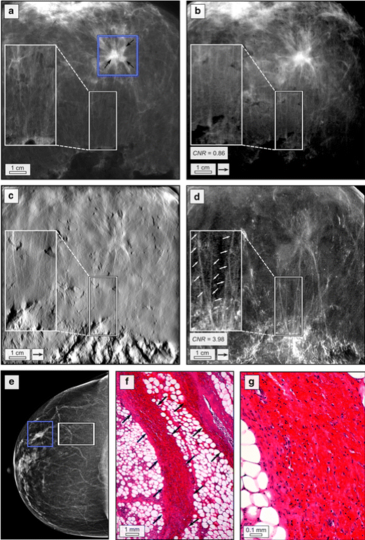
Figure: Dark-field mammography reveals pervasion of breast with partially tumorous soft-tissue strands. Clinical ex vivo mammography in craniocaudal (cc) projection at 29 kVp, 120 mAs and 1.22 mGy average glandular dose (rhodium filter) (a), experimental absorption-contrast mammography (b), phase-contrast mammography (c) and dark-field mammography (d) at 40 kVp, 70 mA and 66 mGy mean glandular dose (per scan direction); in vivo mammography in cc projection at 29 kVp, 105 mAs and1.27 mGy average glandular dose (rhodium filter) (e) of patient 2 in craniocaudal projection; blue rectangles in a and e indicate the trifocal carcinoma; white rectangles in a–e indicate partially infiltrated tissue strands emerging from the tumour; exemplary histological image (haematoxylin–eosin staining) of partially tumour-infiltrated tissue strands (f) and (g) indicated by arrows. The arrows in b–d indicate one-directional measurements.