Sellerer, T., Mechlem, K., Tang, R., Taphorn, K., Pfeiffer, F. and Herzen, J. Dual-energy X-ray dark-field material decomposition. IEEE Transactions on Medical Imaging, 40, 974-985 (2020) [https://doi.org/10.1109/tmi.2020.3043303]
Purpose: we present a novel concept called dual-energy X-ray dark-field material decomposition, which transfers the basic material decomposition approach from attenuation-based dual-energy imaging to the dark-field imaging modality. We develop a physical model and algorithms for dual-energy dark-field material decomposition and evaluate the proposed concept in experimental measurements. Our results suggest that by sampling the energy-dependent dark-field signal with two different X-ray spectra, a decomposition into two different microstructured materials is possible. Similar to dual-energy imaging, the additional microstructure-specific information could be useful for clinical diagnosis.
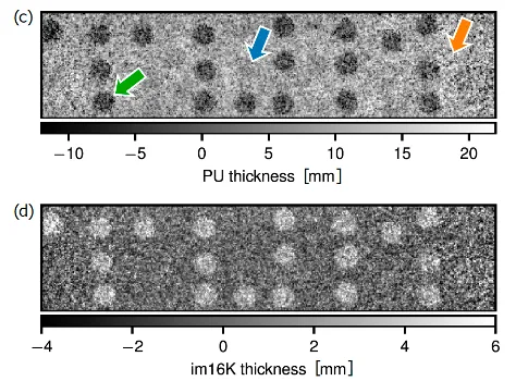
Figure: Sample decomposition. In the conventional dark-field signal acquired with the low (a) and high (b) energy spectrum, the PU and im16K structures cannot be distinguished properly owing to the diminishing contrast. The basis material images, however, provide a clear separation between PU (c) and im16K (d). Further details on the test phantom are given in the text.