Taphorn, K., Mechlem, K., Sellerer, T., De Marco, F., Viermetz, M., Pfeiffer, F., Pfeiffer, D., & Herzen, J. (2021). Direct Differentiation of Pathological Changes in the Human Lung Parenchyma With Grating-Based Spectral X-ray Dark-Field Radiography. IEEE transactions on medical imaging, 40(6), 1568–1578. [https://doi.org/10.1109/tmi.2021.3061253]
Purpose: We investigated the energy-dependent linear diffusion coefficient of simulated lung tissue with different diseases in wave-propagation simulations and validated the results with analytical calculations. Additionally, we modeled spectral X-ray dark-field chest radiography scans to exploit these differences in energy-dependency. The results demonstrate the potential to directly differentiate structural changes in the human lung. Consequently, grating-based spectral X-ray dark-field imaging potentially contributes to the differential diagnosis of structural lung diseases at a clinically relevant dose level.
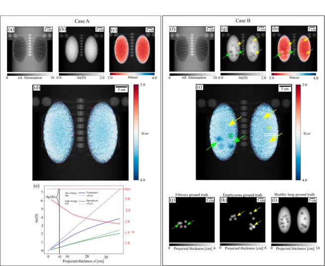
Figure: Spectral X-ray dark-field radiography for two simulated cases. The top row shows the transmission (a,f) and dark-field images (b,g), as well as the overlay of dark-field image and the measured signal ratio qmeas (c,h) from the low and high energy bin. Plot (e) shows the impact of visibility hardening on the measured dark-field signal depending on the projected lung thickness, for the low (solid blue line) and high energy bin (solid green line), in comparison to the expected dark-field signal, calculated with El = 43.5 keV and Eh = 72.0 keV, respectively. Furthermore, the behavior of qmeas is depicted with a solid red line. For increasing sample thickness, qmeas is decreasing and the difference to the expected signal ratio q is increasing. The corrected signal ratio qcor, calculated with eq. (15) is depicted in (d) and (i). Case A represents a healthy phantom. Phantom B had CPFE and qcor demonstrates that the reduction of the dark-field signal in the lower right lobe is related to an increased energy-dependency and fibrosis which is in accordance with the ground truth of fibrotic tissue (j). The ground truths for emphysematous and healthy lung tissue are depicted in (k) and (l), respectively.