Heck, L., Dierolf, M., Jud, C., Eggl, E., Sellerer, T., Mechlem, K., Günther, B., Achterhold, K., Gleich, B., Metz, S., Pfeiffer, D., Kröninger, K and Herzen, J. Contrast-enhanced spectral mammography with a compact synchrotron source. PloS one, 14(10), p.e0222816. (2018). [https://doi.org/10.1371/journal.pone.0222816]
Purpose: We propose to perform another approach for the iodine image calculation based on the dual-energy two-material decomposition method, for which we expect a better quantification of iodine contrast agent. With this approach, further spectral information can be exploited based on the characteristic property of the energy dependency of the linear mass attenuation coefficient.
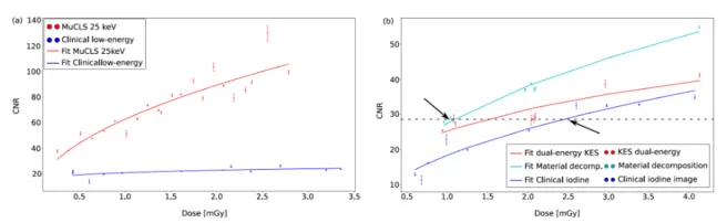
Figure: Dose-dependent overview of the CNR of the iodine solution for (a) the low-energy images and for (b) the iodine images acquired at the MuCLS and with the clinical SenoBright device. Thereby, the dots indicate the results of the measurements at different doses and the lines are the fitted root functions to the data to be able to see a tendency of the CNR with increasing dose. In both subfigures, it is clearly visible that the CNR of the laboratory results is better than of the clinical measurements with the SenoBright system. For the calculation of the iodine image, the two-material decomposition is the method with the highest CNR values. The possibility of reducing the MGD from clinically 2.5 mGy to 1 mGy is indicated by the black arrows and the intersection line in (b). The comparison of KES and material decomposition shows a reduction of 33% from 1.5 mGy down to 1 mGy which underlines the improvement of CESM with the material-decomposition approach.