Hetterich, H., Webber, N., Willner, M., Herzen, J., Birnbacher, L., Hipp, A., Marschner, M., Auweter, S. D., Habbel, C., Schüller, U., Bamberg, F., Ertl-Wagner, B., Pfeiffer, F. and Saam, T. European Radiology 26(9), pp. 3223–3233 (2015). [http://doi.org/10.1007/s00330-015-4143-z]
Purpose: To evaluate the potential of grating-based phase-contrast computed-tomography (gb-PCCT) to classify human carotid and coronary atherosclerotic plaques according to modified American Heart Association (AHA) criteria.
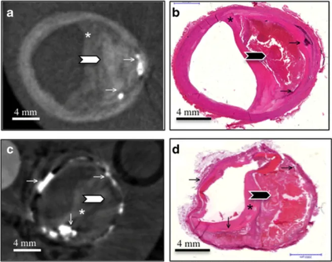
Figure: Example of a type VI lesion in the left common carotid artery (A and B) and right coronary artery (C and D). Axially reconstructed grating-based phase-contrast computed tomography slices (A and C) and corresponding Haematoxylin/Eosin stained histology sections (B and D). Type VI lesions are characterized by a complex plaque with haemorrhage or thrombus and possible surface defect. These atherosclerotic lesions show a large lipid-rich necrotic core with haemorrhage (thick arrows) and overlying thin fibrous cap (*). While the lipid rich areas show little phase shift magnitude, the hemorrhagic areas display a higher intensity level. Several calcifications with high phase shift magnitude can be found in these slices (thin arrows). All these components cannot be visualized with standard attenuation-based imaging.