Willer, K., Fingerle, A. A., Noichl, W., De Marco, F., Frank, M., Urban, T., Schick, R., Gustschin, A., Gleich, B., Herzen, J., Koehler, T., Yaroshenko, A., Pralow, T., Zimmermann, G. S., Renger, B., Sauter, A. P., Pfeiffer, D., Makowski, M. R., Rummeny, E. J., Grenier, P. A. and Pfeiffer, F. X-ray dark-field chest imaging for detection and quantification of emphysema in patients with chronic obstructive pulmonary disease: a diagnostic accuracy study. The Lancet. Digital health, 3(11), e733–e744 (2021). https://doi.org/10.1016/S2589-7500(21)00146-1
Purpose: The novel method of x-ray dark-field chest imaging might fill this gap but has not yet been studied in living humans. Enabling the assessment of microstructural changes in lung parenchyma, this technique presents a more sensitive alternative to conventional chest x-rays, and yet requires only a fraction of the dose applied in CT. We studied the application of this technique to assess pulmonary emphysema in patients with chronic obstructive pulmonary disease (COPD).
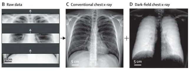
Figure . Examples of raw detector imaging data (B), retrieved conventional chest x-rays (C), and dark-field chest x-rays (D) from the first in-human application