Ferstl, S., Busse, M., Muller, M., Kimm, M. A., Drecoll, E., Burkner, T., Allner, S., Dierolf, M., Pfeiffer, D., Rummeny, E. J., Weichert, W., & Pfeiffer, F. Revealing the Microscopic Structure of Human Renal Cell Carcinoma in Three Dimensions. IEEE transactions on medical imaging, 39(5), 1494–1500 (2020). https://doi.org/10.1109/TMI.2019.2952028
Purpose: We recently developed a laboratory-based method combining nanoscopic X-ray CT with a cytoplasm-specific X-ray stain. Here, we present the application of this method to human RCC biopsies. The NanoCT slices enable pathological characterization of crucial structures by reproducing tissue morphology with a similar detail level as corresponding histological light microscopy images. Beyond that, our data offer deeper insights into the 3D configuration of the tumor. By demonstrating the compatibility of the X-ray stain with standard pathological stains, we highlight the feasibility of integrating staining based NanoCT into the pathological routine.
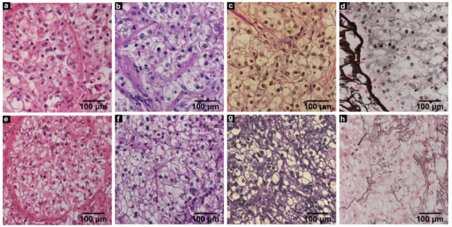
Figure: Comparison of light microscopy images showing histological sections of an RCC (patient B) with different stains.