Gromann, L. B. et al. In-vivo X-ray Dark-Field Chest Radiography of a Pig. Nature Publishing Group 7, 134–7 (2017). [http://doi.org/10.1038/s41598-017-05101-w]
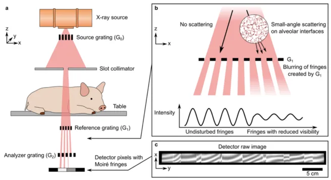
Full-field X-ray dark-field (XDF) chest radiography scanner. (a) Schematic of the prototype. A coarse array of Moiré fringes serves as a reference pattern created by a slight mismatch between the G1 and G2 grating orientation. The anesthetized pig is placed on a sample bed and scanned by a continuous movement. The influence of the sample on the Moir. fringe is used to calculate the XDF images. (b) In case of the lung, millions of micron-sized alveoli (more precisely their air-tissue interfaces) scatter the X-rays, causing a blurring and subsequent decrease of the G1 fringe visibility. (c) Raw detector image with the reference Moir. fringe pattern. Note that the vertical strikes arise from stitching together the borders of neighboring grating tiles and that the scale bar corresponds to the dimensions in the detector plane.