Frank, M., Urban, T., Willer, K., Noichl, W., De Marco, F., Schick, R., Gleich, B., Schegerer, A., Lechel, U., Meyer, P., Mohr, J., Koehler, T., Yaroshenko, A., Maack, I., Pralow, T., Proksa, R., Renger, B., Noël, P., Fingerle, A., Pfeiffer, D., Rummeny, E., Herzen, J. and Pfeiffer, F. Dosimetry on first clinical dark-field chest radiography. Medical physics, 48(10), 6152–6159 (2021). https://doi.org/10.1002/mp.15132
Purpose: The purpose of this study was to evaluate the dose characteristic for patient examinations at the first clinical X-ray dark-field chest radiography system and to determine whether the effective patient dose is within a clinically acceptable dose range.
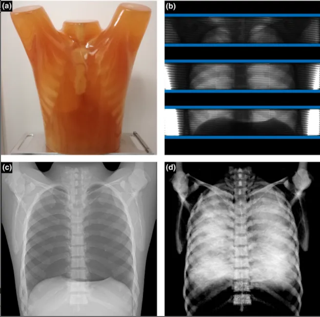
FIGURE
Principle of data acquisition and image processing based on the example of the LUNGMAN phantom (photograph in (a)). During one scan, multiple frames with overlapping boundaries of the active area are acquired with a pulsed X-ray source. Three exemplary, non-overlapping illuminated slot frames are depicted in (b), the slot width is indicated by blue lines. The reconstructed attenuation and dark-field images are shown in (c) and (d), respectively. The most prominent feature in the dark-field image is the lung, which is here modeled by cotton wool placed in the lung cavity