Hellbach, K., Baehr, A., De Marco, F., Willer, K., Gromann, L. B., Herzen, J., Dmochewitz, M., Auweter, S., Fingerle, A. A., Noël, P. B., Rummeny, E. J., Yaroshenko, A., Maack, H.-I., Pralow, T., van der Heijden, H., Wieberneit, N., Proksa, R., Koehler, T., Rindt, K., Schroeter, T. J., Mohr, J., Bamberg, F., Ertl-Wagner, B., Pfeiffer, F. and Reiser, M. F. Scientific Reports 8(1), p. 2602 (2018). [http://doi.org/10.1038/s41598-018-20985-y]
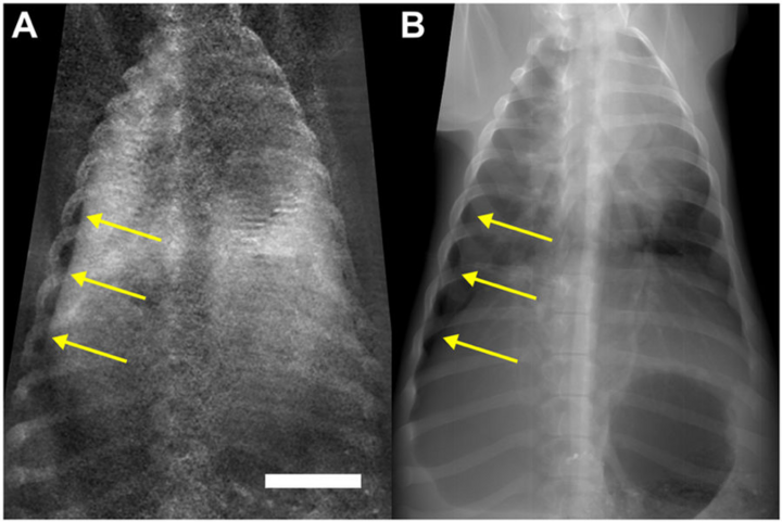
Purpose: The aim of this study was to assess the diagnostic value of x-ray dark-field radiography to detect pneumothoraces in a pig model.
Figure: Unilateral pneumothorax depicted in x-ray dark-field (A) and transmission images (B). Chest radiographs of a pig (in vivo) after a pneumothorax (arrows) was induced on the right side. The transmission image can be compared directly to the corresponding dark-field image. The white scale bar is approximately 5 cm.