
From tissue section to 3D image
New X-ray method materials physics promises advances in histology
An innovative method developed by Dominik John, together with an international team of researchers from TUM BIP (Research Group Biomedical Imaging Physics), Hereon Institute of Materials Physics, and Monash X-ray Imaging Group, centers on a non-destructive technique that combines dyes with 3D X-ray imaging. Using a new algorithm, tissue and dye can be visualized separately in 3D and quantified — opening up new possibilities for research and medicine. The research team presents its study in the journal Advanced Science.
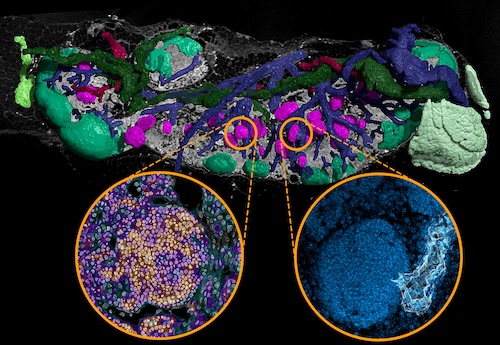
Hidden areas in lymph nodes
How immune stress and age shape lymph nodes: New structures reveal adaptation to chronic stress
Using a high-resolution X-ray technique, an interdisciplinary team from Hereon Institute of Materials Physics and the Research Group of Biomedical Imaging at TUM has revealed novel structures in the lymphatic system of mammals. The 3D images reveal small, previously overlooked clusters of dormant B cells – known as nodules – which are thought to play an important role in the basic readiness of the immune system. The findings expand our understanding of how lymph nodes are structured and how they control immune cells. The study was recently published in the journal Frontiers in Immunology.
More information on Hereon's press release
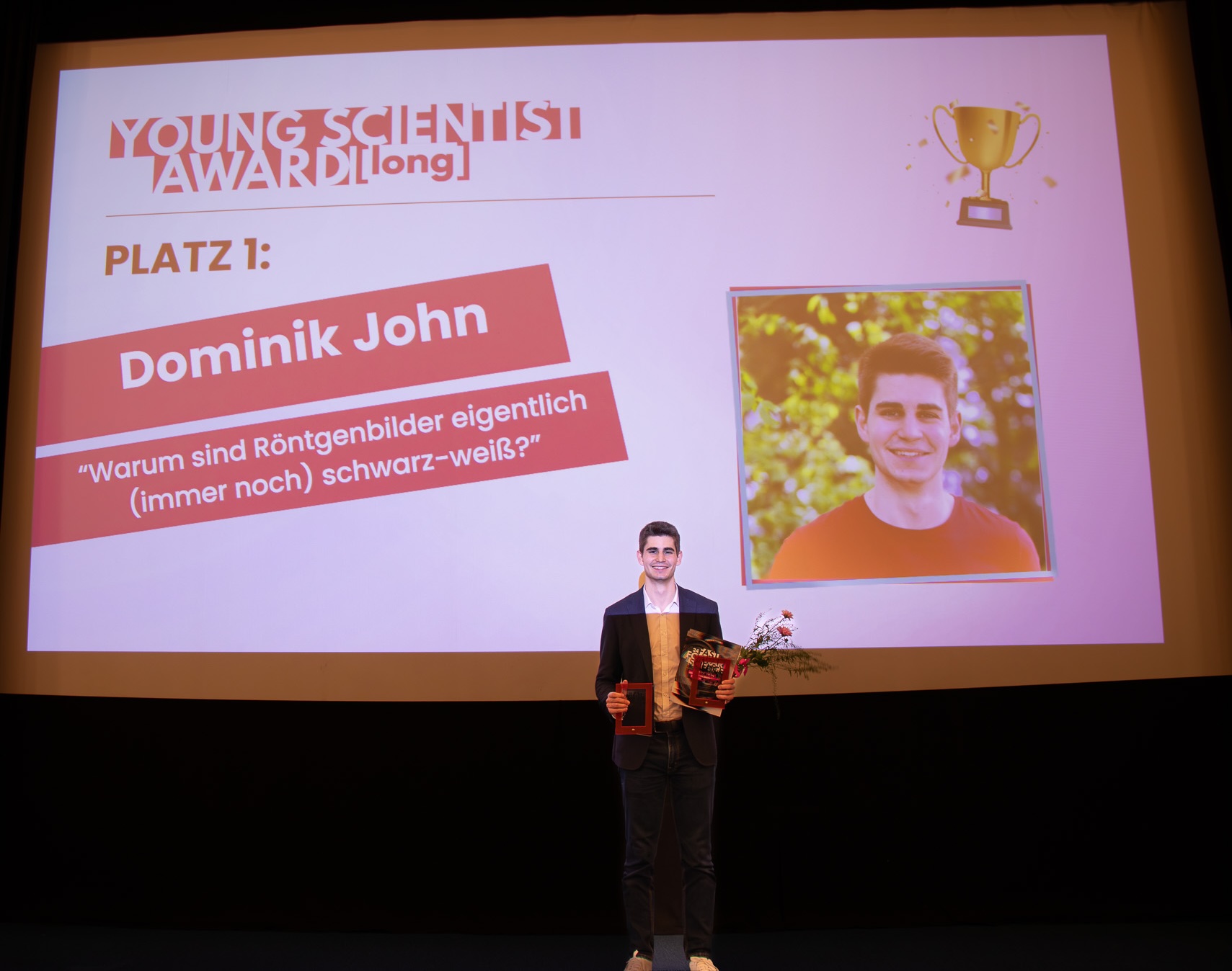

PhD student Dominik John won the first prize in the competition Fast Forward Science 2025 in the category „Young Scientist (long)“
The Fast Forward Science honors young researchers (students, PhD students, or postdocs) who manage to convincingly explain current research with a video more than 3 minutes in length. The Young Scientist award is awarded in collaboration with the team of the Deutscher Zukunftspreis. Dominik John was also the only candidate to simultaneously receive the first prize in a second category, „Best Debut Video“, since it was his first video. It explores the question of why X-ray images are black-and-white and how our modern X-ray research is trying to bring „color“ into the images using new technologies like photon-counting detectors, with the ultimate goal being better diagnostics (e.g. for better detecting cancer). You can find it here: https://www.youtube.com/watch?v=f9gRtqcK1s8
Link to the Fast Forward Science webpage with the annoucements

TUM Press Release 2023 - Three ERC Consolidator Grants for TUM researchers
ERC Consolidator Grant awarded to Prof. Julia Herzen
Prof. Julia Herzen is awarded an ERC consolidator grant for her project DEPICT. She aims to develop a physical model to advance high-resolution X-ray imaging on the micrometer scale and thus be able to determine the composition, distribution, and the amount of individual substances in the samples at such high resolution.

MIBE News 2022 - How contrast agents disperse inside cells
Detailed images of cells with X-ray contrast agents
Contrast agents are often used to improve the imaging of soft tissue in micro-computed tomography (microCT). Now a research team led by the Technical University of Munich (TUM) has investigated how these agents disperse inside cells. Their findings could improve the assessment and further development of contrast agents and might contribute to future medical diagnostics.
TUM Press Release 2022 - Advances in micro-computed tomography
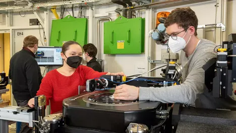
Improved imaging for medicine and material sciences
Researchers in biomedical physics and biology have significantly improved micro-computed tomography, more specifically imaging with phase contrast and high brilliance x-ray radiation. They have developed a new microstructured optical grating and combined it with new analytical algorithms. The new approach makes it possible to depict and analyze the microstructures of samples in greater detail, and to investigate a particularly broad spectrum of samples.